Introduction
The following information is a guide to your upcoming surgery. It describes what is likely to happen at the time of your surgery. This is a guide only and there maybe some individual variation depending on your circumstances and Dr Lawrie will discuss this with you. Please become familiar with this guide so that you can discuss any aspects you wish to with your surgeon, anaesthetist and/or nursing staff.
The Knee Joint
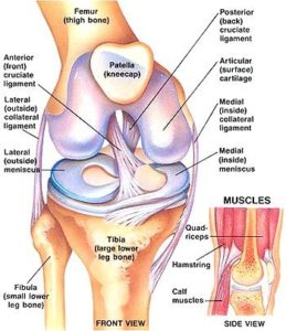
The knee is the largest joint in the body and one of the most easily injured. It works as a complex rotating hinge. It is made up of the lower thigh bone (the femur) the upper end of the shin bone (the tibia) and the kneecap (the patella) which lies at the end of the femur.
There are 4 major ligaments which control the movement and stability of the knee joint. The medial ligament is on the inside of the knee. The lateral ligament and posterolateral corner is on the outside of the knee. These two ligaments control mainly side to side movement.
The anterior cruciate ligament and the posterior cruciate ligament sit one behind the other in the middle of the knee and these are very important ligaments for stability from front to back and rotation. The strong thigh and calf muscles add to strength and stability and also control the movement of the kneecap. The ends of the bones are covered in specialised cartilage called hyaline or articular cartilage which allows for very smooth friction-free movement about the knee. This is the surface that is damaged in osteoarthritis or wear and tear arthritis or inflammatory conditions such as rheumatoid arthritis.
It can also be damaged due to trauma and sports injuries. Sitting on top of the surface of the tibia are two c-shaped cartilages called the meniscus. The meniscus can be thought of as the shock absorber in the knee. They absorb much of the forces that pass through the knee and dampen the shock that is applied to the articular cartilage. They also add some stability to the knee as well as nutrition to the joint surface.
The menisci are often torn with the ACL. Preserving and repairing these cartilages is very important for the long term health of the knee. The knee joint is surrounded by a thin connective tissue layer called synovium that secretes lubricating fluid which reduced the friction to nearly zero in a healthy knee but when this is inflamed it can cause considerable pain and swelling.
About The ACL
The ACL is a strong ligament that sits inside the knee; it runs from the front of the tibia to the back of the femur. It controls front to back stability of the knee as well as rotational stability of the knee. The anterior cruciate ligament is commonly injured insporting injuries and with severe trauma to the knee.
It is much more commonly injured than the stronger posterior cruciate ligament. An anterior cruciate ligament reconstruction is what we commonly referred to as a knee reconstruction. Not everyone who tears their anterior cruciate ligament requires a knee reconstruction.
The ACL is probably not necessary for sedentary activities such as office work, walking and getting up out of a chair, climbing stairs etc. Older patients often don’t require ACL reconstruction after injury. The ACL becomes more important with sporting activities involving sudden changes of direction, contact sports and heavy manual labour. Most ACL injuries are non contact injuries. They typically involve a change of direction with a shift of the body weight over the foot which is planted on the ground. The patient feels a snap and gets immediate pain and is unable to walk on. Typically as the pain and swelling settles down the knee starts to feel good again after a few weeks and the patient is able to walk and perform general day to day activities. However, when they return to the sporting field and do heavy lifting and twisting the knee feels unstable and gives way.
When the knee continues to be unstable it causes damage to the important structures in the knee ( the menisci and articular cartilage) that protect the knee from wear and tear arthritis. The role of ACL reconstruction is to prevent further damage to the knee from ongoing instability, to protect the menisci and articular cartilage and to allow a return to high function. An ACL reconstruction involves using other structures to substitute for the anterior cruciate ligament.
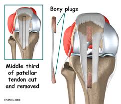
Once the anterior cruciate ligament is torn it does not heal and it cannot be sewn up, this is why a reconstruction and not a repair is necessary. There are a number of reasons to perform an anterior cruciate reconstruction. Typically a graft is made from the hamstring tendons, sometimes part of the patella tendon controlling the kneecap can be used. The patella tendon is typically used if a problem is found intra-operatively with the hamstring tendons or in revision surgery. Other tissues are used more rarely, including donor tissue in revision surgery.
Occasionally an artificial ligament the LARS device may be used. The LARS device should be thought of as experimental surgery. It is only suitable for a very specific type of ACL injury in most patients it is not only unnecessary but not suitable, Dr Lawrie will discuss this with you if needed. There are certain situations when an ACL reconstruction should be done soon after the knee injury. That is if there are associated injuries, especially a meniscal tear that needs to be repaired, then the ACL reconstruction should be performed as soon as possible. This allows the best chance for the meniscus to heal and function normally.
In complex injuries such as high speed road trauma with multiple ligament injuries then early reconstruction will give the best outcome. In the patient who is going to return to a high level of sport and has a very high chance of ongoing instability then they should have their knee reconstructed early to prevent further damage. If the patient is at low risk of further damage to their knee and has no other structures damaged within their knee and they also have a relatively sedentary lifestyle then it is quite reasonable to treat this patient with physiotherapy and allow the muscles about their knee to take over the role of the anterior cruciate ligament.
In patients in their forties or older ACL reconstruction is often unnecessary. In older patients the knee often stiffens up after injury rather than being unstable. Older patients often find the rehabilitation process quite difficult. Once surgery has been decided upon it is of vital importance that the entire rehabilitation course is completed. Many older patients do not return to sport after surgery despite intending too before surgery.
Once surgery has been decided upon it is extremely important that the patient realises there is a lengthy rehabilitation process involved. The most common reason for failure of the graft is if the patient does inappropriate activities with the knee before it is ready. It usually takes 9 months before the knee is ready to return to the sporting field. However, the knee is usually feeling very good by 2-3 months following surgery and it is easy to get bored and think that the knee feels fine so the patient feels they will be right to go back and surf or play football and so forth.
The reconstructed ligament goes through a natural process of changing from a tendon to a ligament which takes around 2 years in total. At 3 months it is actually at its weakest point and by 6-9 months it is strong enough to accept the load on the sporting field. ACL reconstruction surgery is often performed as Day surgery or a single overnight stay.
The patient usually needs crutches for the first 2 weeks with the knee in a splint in slight flexion for the first 2 weeks. The patient can take the knee out of the splint to do some gentle exercises, but the knee is quite sore for the first 2 weeks. From 2-6 weeks the patient can start to weight bear and bend the knee as per the protocol at the end of this information sheet. An exception to this is if a meniscal repair has been performed. Then to protect the meniscus the knee is placed in a range of motion brace set at 0-90° and they will not be allowed to walk on that knee for the first 6 weeks; this is to protect the meniscal repair rather than the graft.
Before Surgery
Pre-operative physiotherapy can be very important. The better conditioned the muscles about the knee are before surgery the better and easier recovery will be. If surgery is performed soon after injury then muscle wasting and deconditioning can be kept to a minimum.
Early surgery also limits further injury to the menisci. The knee is ready for surgery when the inflammation from the injury has settled and full range of motion has returned to the knee. You should stop anti-inflammatory drugs at least five days prior to surgery as they can increase the chance of bleeding (e.g. Voltaren, Naprosyn, Nurofen, Brufen, Feldene, Naprosyn and Indocid) (Celebrex and Mobic are not a problem)
It is extremely important that you have no cuts, scratches or sores on your lower limbs at the time of surgery. If you have any cuts or scratches present then this will increase the chances of infection and your surgery should be postponed until they are well and truly healed. Please contact Dr Lawrie’s rooms well before your surgery day if you notice any of these problems or have any concerns. Postponing the surgery on the day of your operation is very distressing for all concerned. If this can be done ahead of time the theatre time may be used for another patient and resources can be redirected. There is a lot of logistical work that goes into your surgery, this will be lost if the operation is postponed on the day.
On the day of surgery a nurse will inspect your leg for lesions. The nurse will use clippers to remove long hair from the knee and dress the knee with an antiseptic solution. Dr Lawrie will also inspect the leg and mark the appropriate leg to be operated on with an indelible marking pen. You should stop smoking prior to surgery. Smoking increases the risks associated with an anaesthetic and the risk of blood clots (DVT) following surgery.
Admission
You should not eat or drink 6 hours prior to surgery. You should bring with you:
- Personal effects (nightgown or pyjamas, slippers, dressing gown, toiletries) in case you are required to remain in hospital overnight.
- Any medication you are taking
- All relevant x-rays, scans and reports
- Medicare and private health fund membership cards
- This paperwork
Prior to going to the operating room, you will be taken to the Day Surgery Unit and you will:
- Have a shower with an antiseptic soap
- Be dressed in a theatre gown and disposable underwear
- Have the limb inspected and dressed with an antiseptic solution and sterile drape
If you wish Dr Lawrie to speak to a family member after the operation please notify the nursing staff with contact details.
The Procedure
You will be taken into the operating room on a trolley by the nursing staff. The operation will take about 60-90minutes but you may spend up to two and a half to three hours in the theatre complex by the time you wait in the pre-operative area, are prepared from your anaesthetic and then woken up in the recovery area.
The Anaesthetic
The anaesthetist will give you a general anaesthetic. The anaesthetist will discuss this with you prior to the operation.
A drip will be placed in the back of your hand where anaesthetic medicines and antibiotics ca be injected. You will be asked to breathe into an oxygen mask and the drugs to make you fall asleep will be injected through the drip. You will be asleep and not aware of anything further until the operation is over and you have woken up in recovery.
It is not uncommon to have some amnesia prior to or immediately after the surgery due to the anaesthetic drugs.
The Surgery
You will be placed on the operating table lying on your back. A well padded tourniquet will be applied to your thigh and inflated to a safe level just prior to the commencement of surgery. The tourniquet prevents bleeding into the knee and so allows the surgeon to see and perform the surgery easier.
Two to four small one centimetre incisions placed about the knee will be used to gain access to the knee. Through one of the incisions (or portals) the Arthroscope will be introduced. The Arthroscope is like a telescope and is attached to a light source and a video camera. The video camera is attached to a monitor that allows images of your knee to be projected onto the screen.
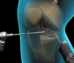
The Arthroscope is also attached to a fluid source (salty water) which constantly flows through the knee. This allows the arthroscope to be manoeuvred through the knee and give a good image on the screen. It also allows any debris or inflammatory chemicals produced within the knee which are causing you pain to be washed from the knee. Through a second portal fine instruments are introduced which are used to correct the injury or disease within your knee.
These instruments include scissors and electronic and manual tissue resecting devices. Suturing devices can also be used. The knee is thoroughly inspected to confirm the diagnosis and to address any other pathology within the knee such as a meniscal tear. At this stage a decision will be made to repair or resect any meniscal pathology. If a meniscal tear is present and is repairable then a number of different suturing techniques may be employed.
This may necessitate a further incision and/or change the post-operative rehabilitation coarse. Menisical preservation is very important for the long term health of the knee. Every effort will be made to repair any mensical tear present. The size position and nature of the tear, the quality of the meniscal tissue itself and the age of the tear determines whether the meniscus can be repaired or has to be removed. The meniscus is usually repaired using an implanted meniscal suture device which has two little barbs connected to a suture and sliding knot which is placed across the tear using the arthroscope.
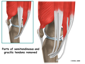
Occasionally an inside out suture is used necessitating an incision on the outside of the knee as well. If the meniscus is not repairable than only the torn part of the meniscus is removed with the remaining meniscus being fashioned into a smooth remnant. The ACL graft is then obtained from one of two donor sites. Either a bone-patellar tendon-bone (BPTB) or Hamstring (HS) graft will be harvested and fashioned to act as the new ACL.
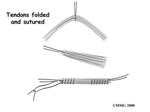
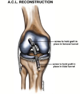
A somewhat larger incision is used to harvest these grafts. Either at the front of the knee for BPTB or on the upper-inner side of the tibia for the HS graft. Hamstring grafts are much more commonly used with BPTB used as a back up if there is a problem with the hamstrings (too small, short weak etc). Two hamstring tendons on the medial side of the knee are removed from there muscle bellies using a special tool. These two tendons are then folded over and sewn together to form the new ACL graft. Small tunnels are drilled in the bone utilising the arthroscope and special jigs to ensure accurate placement of the graft. The graft is inserted into the drill holes and fixed in place with special screws. On occasion supplementary fixation in the form of staples or special Endo-buttons may be used.
The knee is then inspected again with the arthroscope to ensure that the graft is positioned appropriately and functioning properly. The wounds are then sutured closed with dissolvable sutures or staples. On occasion a drain is placed in the wound to prevent excessive swelling. This will be removed within 24 hours. At the end of the procedure your knee will be injected with a combination of local anaesthetic and morphine. A large bandage will be placed around your knee to prevent bleeding and swelling. A knee immobiliser splint will be applied to your knee.
 The Recovery
The Anaesthetist will reverse the anaesthetic and you will wake up in the recovery room where the nurse will check your observations and movement in your feet. Once you are sufficiently awake you will return to the Surgical Ward.
After Surgery
Dr Lawrie will see you and explain the operative findings to you. A copy of the operative report will be given to you and sent to your local doctor. Once you are well and truly awake you will be visited by the physiotherapist who will instruct you on a series of exercises to get the best result from your operation.
This will be targeted at strengthening specific muscle groups so as to protect the knee from damage in the early post-operative stages and to get the knee on its way to moving normally again. A specific post-operative physiotherapy programme is attached. Overnight you will be given morphine or morphine like medication for pain.
This may be in the form of a PCA (Patient Controlled Analgesia). On discharge you will have tablets available to take home with you (Panadiene forte/ Tramal/ Digesic/Endone). You may also be given an anti-inflammatory type medication. You can travel home in a car, but can not drive. The night of surgery your knee should be reasonably comfortable due to the local anaesthetic and morphine within the knee. This typically wears off after 12-18 hours. It is normal to experience some discomfort the following day as this wears off.
When You Get Home
The amount of post-operative discomfort can be quite variable and is dependant on the type and amount of surgery that was performed. It is quite normal for the knee to be painful and stiff for the first two weeks. It often takes up to six weeks before the knee is feeling truly comfortable. If you experience severe pain or swelling or have any worries contact Dr Lawrie or and/or your local doctor. The bulky bandage should remain on your knee for three days to help with postoperative swelling. After this the bandage can be removed and small Primapore type dressings can be applied. A neoprene (wet suit material) knee sleeve is very useful to relieve swelling and can be worn following removal of bandages.
It is very important that the surgical wounds are kept clean and dry whilst they are healing. The wounds are generally healed after 12 days. If sutures are present these should be removed at day 12-14 post-operatively. Steri-strips should be allowed to full off on their own after day 12. A strip of Hypafix tape should be applied to the hamstring wound on the shin. This should be changed on a weekly basis for 3 months. This reduces tension on the wound helping with wound healing.
You may drive a vehicle when you can walk comfortably without crutches and are not taking pain killers that may cause drowsiness. It is a good idea to consult with your insurance company about when you can return to driving. You will be seen for follow-up in Dr Lawrie’s rooms after the surgery. The timing of which will be decided on at the time of surgery. We will again discuss the findings at the time of surgery, what surgery was performed and what the future holds for your knee.
Commonly Asked Questions
Will my surgery be successful?
ACL reconstruction has a high success rate. Graft failure occurs in 10-15% of patients over a ten year follow-up period
Will I need further surgery?
It is unclear whether ACL reconstruction will decrease the need for further surgery in the long term. If we can prevent further injury to the knee and preserve the menisci and joint surface the knee should do much better in the long term. At the time of injury it is common to suffer a bone bruise to the femur. This may result in early onset osteoarthritis in the long term. However it is understood that successful ACL reconstruction will protect the knee from further damage such as to the menisci or shock absorbing cartilages
When can I drive?
When you are walking without crutches, have good quadriceps function and are not needing pain killers which may cause sedation
When can I return to work?
If a sedentary job then at about two weeks one can return to work. Manual labour puts quite a strain on the graft and one can usually return to light duties in 2-3 months and then full duties at around 4-6 months
When can I return to sport?
This will depend on the type of sport, level of sport and your post-operative recovery. Return to high level, organised contact sport generally takes about twelve months. Playing sport non-competitively or training may be possible at 6-9 months but is assessed on an individual basis.
Post-Operative Rehabilitation For ACL Reconstruction
Day 1
Surgery
Day 1
Home in splint
Can take splint off for exercises
Touch weight-bear with crutches
At week 1
Gentle exercises
Encourage calf movement
Partial weight-bear by end of week
At week 2
Reviewed by Dr Lawrie
Begin regular physiotherapy
Remove splint
Should have 90° bend
Fully weight-bear
Co-contractions, Straight leg raise with no resistance
At week 3
Aim to walk independently
Ensure full extension
At week 4
Aim for 0-100° bend with full weight-bear
Quads and hamstrings biofeedback/stimulation
Prone hangs if not full extension 10cm step ups, calf raises
Partial squats to 45° bilaterally bike (no tension), gentle wobble board
At week 6
Add swimming – gentle only – no flicking
At week 10
Progress exercises and add resistance.
ALL QUADS EXERCISES MUST BE CLOSED CHAIN
Aim 0-130°
Add mini-tramp, wobble board, lunges and slide board
At 4 months
Begin dynamic activity
Increase speed and agility
Graduate all exercises to develop strength
Begin light jogging
At 5 months
Progress running and early sport activity
Begin Fig8, direction changes, slopes
Begin sport specific exercises and individual skills
Protect ACL with a knee sleeve or taping
At 9 months
If all parameters are good and confident of ability – return gradually to full activity
If meniscal repair performed:
1st 6 weeks in ROM brace limiting ROM 0-90° and non weight bearing first 6 weeks to protect meniscal repair
Encourage ROM especially full extension in brace, pushing quadriceps, co-contractions and straight leg raises